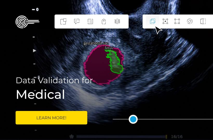
Image Classification for Medical Image Analysis
Radiologists must interpret one image every 3-4 seconds to meet clinical demands. This fact underscores the critical need for advanced medical image classification technologies. As medical imaging volumes surge, AI in healthcare is becoming vital.
Computer vision and disease detection algorithms are transforming medical image analysis. These technologies are not only keeping up with human experts but are often outperforming them in diagnostic accuracy across various imaging modalities.
The field of medical image classification is undergoing rapid evolution. Last year, researchers published four groundbreaking articles on deep learning in medical imaging. This marks a significant increase from just one article in 2014, reflecting the exponential growth in this crucial healthcare technology domain.
Key Takeaways
- Radiologists face immense pressure, interpreting an image every 3-4 seconds
- AI in healthcare is matching or exceeding human expert accuracy in image analysis
- Deep learning models are transforming medical image classification
- Research in this field has grown significantly since 2014
- AI technologies cover various aspects, including tumor identification and fracture detection

Understanding Medical Image Classification
Medical image classification is a vital component of modern healthcare. It involves the analysis of various medical images to support diagnosis and treatment planning. The field has witnessed significant advancements, thanks to technology. These advancements have improved diagnostic accuracy and helped manage the workload of radiologists.
Definition and Importance in Healthcare
Medical image classification is the process of categorizing medical images based on their features. This technique is crucial for early disease detection and creating personalized treatment plans. A recent review of 425 peer-reviewed articles from PubMed and Web of Science underscores the growing significance of this field in healthcare.
Types of Medical Images
Several medical imaging techniques are employed in diagnosis:
- X-rays: Commonly used for bone fractures and chest examinations
- MRI (Magnetic Resonance Imaging): Ideal for soft tissue imaging
- CT (Computed Tomography) Scans: Provide detailed cross-sectional images
Challenges in Manual Image Interpretation
Despite advancements, manual image interpretation faces several challenges:
- High volume of images compared to available radiologists
- Time constraints leading to potential delays in diagnosis
- Risk of human error due to fatigue or cognitive overload
- Need for expert knowledge in specialized areas
These challenges highlight the need for automated systems in medical imaging workflows. Such systems can support radiologists and improve patient care.
| Model Type | Number of Studies | Most Popular Model |
|---|---|---|
| Multiple Models | 57 | Varied |
| Deep Models | 33 | Inception (26 studies) |
| Shallow Models | 24 | Not specified |
The field of medical imaging is evolving rapidly. Integrating advanced technologies like deep learning and transfer learning is becoming increasingly important. These technologies enhance image interpretation and reduce the workload of radiologists.
The Role of Deep Learning in Medical Imaging
Deep learning has revolutionized medical image analysis, changing the way healthcare professionals diagnose and treat patients. This technology employs neural networks to analyze complex medical images with unmatched accuracy and speed.
Advancements in Computer Vision for Healthcare
The growth of deep learning in medical imaging has been swift and profound. Since its inception in 2015, it has become a key trend in analyzing medical images. Neural networks now excel at tasks such as detecting microcalcifications in mammograms and segmenting brain tumors in MRI scans.
Comparing Deep Learning to Traditional Methods
Deep learning surpasses traditional computer-aided detection (CAD) systems in several aspects:
- Accuracy: Deep learning models show higher sensitivity and specificity in detecting abnormalities.
- Speed: AI diagnostics can analyze images much faster than human radiologists.
- Consistency: Neural networks deliver consistent results, reducing human error.
Potential Impact on Diagnostic Accuracy and Efficiency
The impact of deep learning on medical imaging is significant:
| Application | Accuracy | Improvement |
|---|---|---|
| Brain Tumor Segmentation | 91.4% | Increased precision in tumor boundary detection |
| Pulmonary Nodule Detection | 90% | Earlier detection of lung cancer |
| Multiple Sclerosis Lesion Segmentation | 82% | Better tracking of disease progression |
These advancements in medical image analysis using deep learning are setting the stage for more accurate diagnoses and tailored treatment plans. As this technology advances, it holds the promise of improving patient care and streamlining healthcare processes.
Convolutional Neural Networks (CNNs) in Medical Image Analysis
CNNs have transformed medical image classification, providing a robust deep learning framework for extracting features. They excel in dissecting complex visual data, crucial for diagnosing conditions such as pneumonia from chest X-rays.
Their prowess in medical image analysis is clear, often surpassing human experts. For example, CheXNet, a 121-layer CNN, outperformed four radiologists in diagnosing pneumonia from over 100,000 chest X-rays. This milestone in medical image classification highlights deep learning's potential in healthcare.
CNNs automatically discern and extract features from images, obviating the need for manual feature engineering. This feature makes them ideal for tasks such as detecting abnormalities, classifying diseases, and diagnosing in medical imaging.
The architecture of a CNN typically includes:
- Input layer
- Convolutional layers
- Activation layers
- Pooling layers
- Fully connected layers
- Output layer
Researchers are now exploring hybrid structures that combine CNNs with technologies like transformers and GANs to boost performance in medical image segmentation and classification. The choice of activation function is critical for optimizing CNN performance, with selections based on empirical evaluation and task-specific requirements.
| CNN Application | Performance |
|---|---|
| Pneumonia Detection | Outperformed human radiologists |
| Chest X-ray Classification | Improved accuracy with data augmentation |
| Interstitial Lung Disease (ILD) Pattern Classification | Customized CNN with shallow convolution layer |
As CNNs evolve, they promise to elevate diagnostic precision and efficiency in medical image analysis, potentially transforming healthcare delivery.
Self-Supervised Learning
Self-supervised learning is transforming medical AI, tackling the hurdle of limited labeled data in healthcare. This method enables machines to learn from vast, unlabeled data, thus reducing the need for costly, time-consuming manual annotations.
Overcoming Limited Labeled Data Challenges
In medical fields, gathering labeled datasets is challenging due to privacy issues and the necessity for expert annotations. Self-supervised learning addresses this by allowing AI models to extract valuable features from unlabeled data. This technique has shown significant success in medical image classification tasks.
A self-supervised approach to training medical image classifiers can dramatically reduce the number of labeled images needed to train the model compared to the typical supervised approach.
Improving Model Generalization Across Institutions
Self-supervised learning enhances model generalization, enabling AI systems to excel across various healthcare institutions. By learning from diverse, unlabeled datasets, these models develop robust features suitable for numerous medical scenarios.
Types of Self-Supervised Learning Strategies
Several self-supervised learning strategies have emerged as effective AI training strategies for medical image analysis:
- Contrastive learning: Models learn to distinguish between similar and different images
- Generative approaches: AI systems reconstruct or predict parts of medical images
- Self-prediction methods: Models forecast future frames in time-series medical data
| Strategy | Description | Application in Medical AI |
|---|---|---|
| Contrastive learning | Distinguishing similar vs. different images | Enhancing disease detection accuracy |
| Generative approaches | Reconstructing or predicting image parts | Improving medical image segmentation |
| Self-prediction methods | Forecasting future frames in time-series data | Analyzing medical video sequences |
As self-supervised learning advances, it holds the promise of unlocking new possibilities in medical AI. This will lead to improved diagnosis accuracy and enhanced patient care through more efficient, adaptable models.
Image Classification for Medical Image Analysis
CNNs are adept at extracting features from images in a hierarchical manner. Lower layers focus on basic elements, while higher layers interpret complex semantic aspects. This hierarchical structure makes CNNs particularly suitable for medical image analysis, where accurate diagnosis hinges on subtle details.
The impact of these techniques is profound:
- A study utilizing 28,378 multi-modal medical images achieved a classification accuracy of 98.61%.
- Deep learning algorithms have the potential to surpass human experts in certain diagnostic tasks.
- Various CNN architectures, including VGGNet, ResNet, and DenseNet, have shown remarkable results in medical imaging applications.
However, challenges persist. Manual annotation of medical images is time-consuming and error-prone. This underscores the necessity for more automated, precise methods in image classification. Researchers and clinicians are now exploring CNN applications for segmentation, abnormality detection, and disease classification to tackle these challenges.
As the field advances, we're witnessing the development of improved CNN structures that incorporate other algorithms like transformers and generative adversarial networks. These hybrid models are promising in enhancing medical image segmentation and classification. They are expanding the possibilities in diagnostic imaging.
Transfer Learning in Medical Image Classification
Transfer learning has transformed medical image classification. It uses pre-trained models to handle complex medical tasks with limited data. By adapting models trained on large datasets like ImageNet, researchers can fine-tune them for specific medical applications.
Leveraging Pre-trained Models for Medical Tasks
Pre-trained models provide a robust foundation for medical image analysis. Initially trained on diverse image datasets, these models can be tailored to various medical imaging modalities. Research indicates that transfer learning significantly boosts performance across different architectures. Smaller models often see the most improvement.
Fine-tuning Strategies for Optimal Performance
Fine-tuning pre-trained models is essential for achieving peak performance in medical image classification. This process involves tweaking the model's parameters to align with specific medical datasets. Strategies include:
- Retraining specific layers
- Adjusting network complexity
- Truncating final blocks to reduce model size without compromising performance
Data Augmentation Techniques for Medical Images
Data augmentation is vital in medical image classification, especially with limited datasets. It involves modifying existing images to increase the training set, thus boosting AI model training capabilities.
Medical image datasets often struggle with scarcity, privacy issues, and labeling challenges. These problems can result in bias, overfitting, and inaccurate results. Data augmentation techniques help by enhancing deep network architectures' learning and generalization abilities.
Researchers have pinpointed 65 distinct data augmentation techniques for medical imaging. These techniques span various categories, including spatial transformation, color adjustment, noise addition, and data mixing. Some methods need manual parameter setting, while others automate based on task demands.
| Augmentation Category | Examples | Benefits |
|---|---|---|
| Spatial transformation | Rotation, flipping, scaling | Improves model robustness |
| Color adjustment | Brightness, contrast changes | Enhances feature recognition |
| Noise addition | Gaussian noise, salt-and-pepper noise | Increases model resilience |
| Data mixing | Image blending, mosaic mixing | Expands dataset diversity |
Data augmentation techniques bring significant benefits to medical image analysis. They reduce overfitting, improve model generalization, and boost performance across imaging modalities. By addressing class imbalance and domain shift in transfer learning, these methods lead to more accurate and robust AI models for medical image classification.
Capsule Networks: A Novel Approach to Medical Image Classification
Capsule networks are a groundbreaking neural network architecture that's transforming medical image analysis. They bring unique benefits to small datasets, especially in tasks like brain tumor detection.
Understanding Capsule Network Architecture
Capsule networks, or CapsNets, overcome traditional CNNs' limitations by maintaining spatial relationships between features. This is essential for precise medical image interpretation, particularly with complex anatomical structures.
Advantages Over Traditional CNNs for Small Datasets
CapsNets stand out in small dataset performance, a frequent hurdle in medical imaging. They achieve high accuracy with fewer training examples, making them perfect for specialized medical tasks where large datasets are scarce.
- Preserves location information and part-whole relationships
- Provides translation equivariance
- Captures richer contextual patterns
Performance in Brain Tumor Detection
In brain tumor detection, capsule networks have shown impressive outcomes. Accuracy rates of up to 86.56% have been reported, highlighting their potential to boost diagnostic capabilities in neurology.
| Model | Accuracy | Parameter Count |
|---|---|---|
| AOC-CapsNet | State-of-the-art in 2/7 tasks | Significantly reduced |
| SegCaps | State-of-the-art | <5% of popular networks |
| Deep Conv-Deconv CapsNet | High performance | ~1,474,560 parameters |
Capsule networks efficiently tackle complex medical imaging tasks, positioning them as key players in advancing AI-powered medical diagnostics.
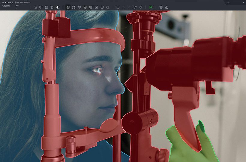
Challenges in Medical Image Classification
Medical image classification encounters distinct hurdles within the healthcare technology domain. The advent of deep learning has transformed disease detection. However, it's essential to confront the obstacles that impede progress in this sector.
Data Privacy and Ethical Concerns
Data privacy in medical image classification is a critical issue. Regulations safeguard patient information, complicating the sharing and accessing of large datasets. This constrains the development of advanced AI for diagnostic and treatment planning purposes.
Need for Expert Annotations
For training precise models, expert annotations are indispensable. Yet, this process is both time-consuming and costly. The dearth of annotated medical images significantly hampers the creation of dependable classification systems.
Dealing with Small and Imbalanced Datasets
Medical imaging datasets often face challenges due to their small and imbalanced nature. Such datasets can result in biased predictions and overfitting. This problem is especially acute in rare diseases with limited sample sizes.
| Challenge | Impact | Potential Solution |
|---|---|---|
| Data Privacy | Limited data sharing | Federated learning |
| Expert Annotations | Time and cost intensive | Semi-supervised learning |
| Imbalanced Datasets | Biased model predictions | Data augmentation techniques |
Ensuring ethical AI in healthcare is vital, balancing innovation with patient rights. With cloud computing in healthcare expected to hit $89 billion by 2027, addressing these challenges is key. It's essential for advancing medical image classification while upholding ethical standards.
Performance Metrics and Evaluation in Medical Image Classification
Evaluating medical image classification models demands a thorough approach. Accuracy is crucial but not the sole metric to focus on. Metrics like sensitivity, specificity, F1 score, and AUC-ROC offer a fuller view of model performance in medical settings.
Accuracy metrics can be deceptive, particularly with imbalanced datasets prevalent in medical imaging. A model might show high accuracy by consistently predicting the majority class, yet miss rare conditions. Therefore, AI benchmarking must encompass a variety of performance indicators.
Diagnostic performance is paramount in healthcare. A study by Litjens et al. from 2012 to 2017 underscored the significance of thorough evaluation methods in deep learning for medical image analysis. They discovered that models with superior sensitivity and specificity generally exhibited better diagnostic accuracy in real-world scenarios.
Comparing AI models to human experts necessitates standardized metrics. Taha and Hanbury introduced metrics for evaluating 3D medical image segmentation, now widely accepted. These metrics facilitate equitable comparisons between AI solutions and human performance.
| Metric | Description | Importance |
|---|---|---|
| Sensitivity | True positive rate | Crucial for detecting diseases |
| Specificity | True negative rate | Important for ruling out conditions |
| F1 Score | Harmonic mean of precision and recall | Balanced measure of performance |
| AUC-ROC | Model's ability to distinguish between classes | Overall classification performance |
The field of medical image analysis is continually evolving, so are the methods for evaluating models. Müller et al. introduced MISeval in 2022, a comprehensive metric library for evaluating medical image segmentation. This tool standardizes evaluation, simplifying the comparison of different models and techniques across various studies.
Future Directions in AI-Powered Medical Image Analysis
AI is increasingly playing a pivotal role in clinical decision support systems. With 14% of reviewed papers highlighting AI's impact on healthcare, the focus is on integrating it smoothly into daily medical routines. This integration aims to enhance human expertise, leading to more efficient and accurate diagnoses.
Multimodal Learning and Fusion Techniques
Multimodal learning is transforming medical image analysis. By fusing various data types, including patient history and genomics, with imaging data, AI systems can deliver more comprehensive insights. Vision transformers in lung cancer diagnosis have boosted prognosis accuracy by 20%. This highlights the strength of advanced AI techniques in complex medical scenarios.
Explainable AI for Medical Imaging
The drive towards explainable AI is essential for establishing trust in AI-generated results. With an average 85% efficacy rate in disease diagnosis, ensuring transparency in AI decision-making is crucial. This development aims to create AI systems that not only perform well but also provide clear explanations for their conclusions. This enables clinicians to make informed decisions with confidence.
FAQ
What is Medical Image Classification?
Medical Image Classification is a process where medical images like X-rays, MRI scans, and CT scans are sorted into different groups. This is done using computer algorithms to automatically identify and categorize images. It aids in making diagnoses, planning treatments, and monitoring diseases.
Why is Medical Image Classification important in healthcare?
It's vital in modern medicine for making diagnostic decisions and planning treatments. With the growing workload for radiologists, who now analyze one image every 3-4 seconds, it helps manage the increasing demands.
How does Deep Learning contribute to Medical Image Classification?
Deep learning models, especially Convolutional Neural Networks (CNNs), have reached human-level accuracy in various medical fields and imaging types. This technology promises to enhance the efficiency and accuracy of medical image interpretation.
What are the advantages of Convolutional Neural Networks (CNNs) in Medical Image Analysis?
CNNs provide an end-to-end solution, automatically learning features without manual engineering. They've shown significant performance in medical imaging tasks, sometimes even surpassing human experts, like in detecting pneumonia from chest X-rays.
How can Self-Supervised Learning help in Medical Image Classification?
Self-Supervised Learning (SSL) offers a solution to the challenge of annotating large medical image datasets. It enables the creation of generalist models that can be tailored for various tasks without needing extensive labeled data, potentially reducing development costs and time.
What are some traditional techniques used in Image Classification for Medical Image Analysis?
Traditional methods include Support Vector Machines (SVMs) with feature descriptors like Scale-Invariant Feature Transform (SIFT) or Oriented FAST and Rotated BRIEF (ORB). These have been effective for classifying medical images.
How does Transfer Learning benefit Medical Image Classification?
Transfer learning is beneficial for medical image classification, especially with limited data. It uses models pre-trained on large datasets like ImageNet and fine-tunes them for specific medical tasks, achieving high accuracy with small datasets.
Why is Data Augmentation important in Medical Image Classification?
Data augmentation is crucial for expanding small medical image datasets. It creates new versions of existing images, significantly boosting model performance and improving generalization.
What are Capsule Networks, and how are they used in Medical Image Classification?
Capsule Networks are a new type of neural network architecture that have shown promising results on small medical imaging datasets, like brain tumor detection. They offer advantages over traditional CNNs in handling small datasets and preserving spatial feature relationships, essential for medical image analysis.
What are some challenges faced in Medical Image Classification?
Challenges include data privacy concerns, the need for expert annotations, and the prevalence of small and imbalanced datasets. These issues require innovative solutions in data handling, model development, and ethical considerations in AI-powered medical image analysis.
How are Medical Image Classification models evaluated?
Models are evaluated using performance metrics like sensitivity, specificity, F1 score, and AUC-ROC. Comparing AI model performance with human expert benchmarks is also crucial for evaluation.
What are some future directions in AI-Powered Medical Image Analysis?
Future directions include integrating AI with clinical workflows, combining different medical data types, and developing explainable AI. These advancements aim to create more robust, versatile, and trustworthy AI systems that can significantly improve patient care and diagnostic accuracy.